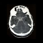
The Arnold-Chiari Malformation, also known as tonsillar ectopia or tonsillar herniation, is a cerebral malfunction where the cerebellum, the area of the brain responsible for muscular movement and coordination descends  through the foramen magnum, invading the spinal cord (cervical vertebral canal). 

This is a malformation that may exist at birth or, which may develop over time. Sometimes, the Arnold-Chiari malformation occurs due to lack of space or due to a malformation at the back of the skull. .

There are four degrees of Arnold-Chiari malformation depending on the extension of the tonsillar descent and the morphological consequences (Grades I, II, III and IV). It is estimated that the incidence of Grade I of Arnold-Chiari malformation is between 0.6 and 0.9 %, with a predominance of 2:3 men over women. The diagnosis would be made between the ages of 30 and 40. The incidence is 0.001% in the case of Grade II of Arnold-Chiari malformation or Chiari II.

We have already mentioned grade I and II while grades III and IV include an important cerebellar hypoplasia, which has a more negative effect on the malformation.  We might say that grade I corresponds to the adult while the other grades are part of malformation in a child.

Symptoms


  • Suboccipital headache

  • Ocular Semiology 

  • Otoneurological symptoms 

  • Sensory and motor deficits

  • Ataxia

  • Dysphagia

  • Damage to cranial nerves

  • Respiratory arrest

  • Sudden death

  • Medulla Disorders

Diagnosis

Cranial Nuclear Magnetic Resonance Imaging (MRI)

The Cranial Magnetic Resonance test is determined for the diagnosis of Arnold-Chiari. This non-invasive test allows us to assess the exact measurement of the tonsillar descent in relation to the data provided by the cisternal and bone image. Typically a lower limit defined at 5mm, below the amount at which the tonsils should be, must be defined to consider the existence of a Chiari malformation I. In the Chiari II, we can find evidence of  the deformity of the medulla oblongata, a fusion of the quadrigeminal sheet intermediate mass dilated elongation of the medulla oblongata and insertion below the tentorium of the cerebellum. Finally, the evaluation and analysis of the dynamic flow image obtained by MRI, allows the  physiopathological knowledge which causes and accompanies the malformation,  having a decisive influence on the design of a therapeutic strategy.

Treatment

The therapy is based on surgical treatment because there is no evidence that medical treatment interrupts the process .

There are various surgical treatment possibilities described. All of them are designed to decompress the cerebellar tonsils and restore the normal flow of fluid between the cranial and spinal compartments.

To do this a multitude of strategies have been proposed, all contemplating these basic decompression principles. Based on the classic posterior fossa craniotomy and opening the dura mater, including the removal of the tonsils.

In our opinion, and given that the aim is the decompression of the cerebellar tonsils, in order to permeabilize cisternal movement to the foramen magnum level, the majority of times, the duraplasty enlargement does not appear to be necessary. The simple craniotomy restricted to the foramen magnum, in order to increase the calibre, is always accompanied by the meticulous extirpation of the abutment, the annular ligament and constrictor, which is situated below the occipital bone,  which will be sufficient to obtain the desired result.


Blogs barnaclínic+