27 July, 2020
What are Skull Base Chordomas and what is its treatment?

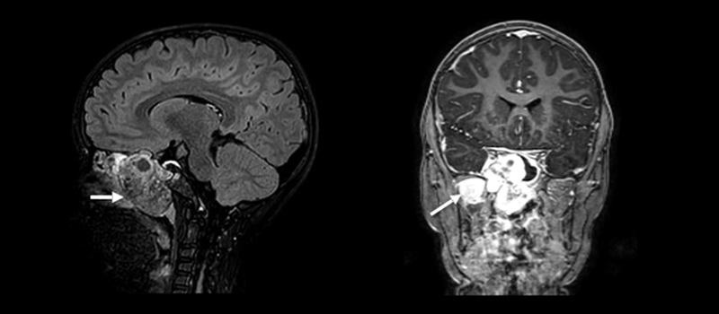
Chordoma is a rare type of tumor that occurs in the bones of the skull base and spine. These tumors arise from the remnants of the notochord, a flexible, rod-like structure that provides support to the developing embryo. Chordomas are complex tumors to treat due to the involvement of critical structures such as the brainstem, spinal cord, and important nerves and arteries. About 30 percent of all chordomas are form within the center of the head in an area called the skull base – usually in a bone called the clivus. Skull base chordomas are sometimes considered brain tumors because they can grow toward the brain.
IT MAY INTEREST YOU…
· What are skull base tumors?
· Brain tumors: symptoms, diagnosis and treatment
Chordoma Prevalence
One in one million people per year is diagnosed of a chordoma. In gross numbers, 300 patients are diagnosed with chordoma each year in the United States, and about 700 in all of Europe.
Most of the patients are diagnosed in their 50s and 60s, but it can occur at any age. Skull base chordomas occur more frequently in younger patients, while spinal chordomas are more common later in life. Chordoma affects almost the double of men than women. While chordoma can run in families, this is very rare.
Probable causes of Skull Base Chordomas
Chordoma tumors develop from cells of a tissue called the notochord. The notochord dissolves when the fetus is about 8 weeks old, but some notochord cells remain in the bones of the spine and skull base. Very rarely, these cells start replicating forming the so called chordoma. What causes notochord cells to become cancerous in some people is not fully known, but researchers are working to found the answers.
Chordoma Classification
Chordomas al classified based on how they look under de microscope:
-
Conventional (or classic) chordoma
Conventional (or classic) chordoma is the most frequent form of chordoma. It is composed of a distinctive cell type that resembles notochordal cells and can have areas of chondroid appearance.
-
Chondroid chordoma
Chondroid chordoma is a term more commonly used in those chordomas with a chondroid appearance. In the past was difficult to distinguish conventional chordoma from chondrosarcoma, but now a days This is no longer a problem because brachyury is expressed in nearly all conventional chordomas, and other cartilaginous tumors like chondrosarcoma do not express brachyury.
-
Poorly differentiated chordoma
Poorly differentiated chordoma is a recently identified subtype. It can be more aggressive and faster growing than conventional chordoma, and is more common in pediatric and young adult patients, as well as in skull base and cervical patients. The diagnosis is based primarily because nearly all poorly differentiated chordomas are INI1-negative, meaning they do not express the INI1 protein.
-
Dedifferentiated chordoma
Dedifferentiated chordoma is more aggressive and generally grows faster than the other types of chordoma, and is more likely to metastasize than conventional chordoma. It can also have loss of the INI1 gene, but this is not frequent. This type of chordoma is uncommon, occurring in only about 5 percent of patients, and is more common in pediatric patients.
Skull Base Chordomas treatment
Today, the optimal treatment for skull base chordoma is still under debate. The objective is to offer the highest resection index, minimizing intra- and post-operative morbidity and mortality. The development of the endonasal endoscopic technique during the last 15 years has managed to equalize the classic degrees of resection (using open surgery and a microscope) and even exceed them, also associating a lower complication rate.
Our barnaclínic+ Skull Base Unit has extensive experience in the management of skull base chordoma, using an extended endonasal endoscope (AEEE) approach for their surgical treatment, carried out by the surgeons Dr. Joaquim Enseñat, Dr. Cristobal Langdon and Dr. Isam Alobid.


